Explanation
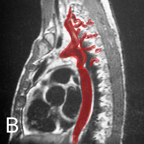
The small structures outlined in red are the intercostal arteries, which come off the aorta (the larger structure outlined in red) at each level and travel just inferior to each rib. Each intercostal artery travels together with an intercostal vein and an intercostal nerve.
There are most commonly 9 pairs of intercostal arteries that arise from the aorta, distributed to the T3-T11 levels. Because the T12 branch is not between two ribs, it is called the subcostal artery. The arteries that run between T1-T2 and T2-T3 most often arise from the costocervical trunk as the supreme (or highest, or superior) intercostal artery. This is somewhat variable, and the arteries to these upper levels can arise from the aorta or from other vessels such as the vertebral artery.
Image A is an unlabeled image from a T1-weighted sagittal MR of a patient with abnormalities of the intercostal arteries.
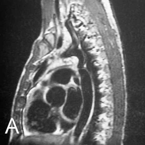
Image B shows the abnormal aorta, outlined in red, which has a marked narrowing just below the level of the ductus: a coarctation. In this condition, the intercostal arteries may become markedly enlarged as they serve as a collateral pathway for blood to get past the area of narrowing, as shown here.
Tidak ada komentar:
Posting Komentar