Rabu, 23 Februari 2011

MMed Anatomy, Spotter

26 Jan Spotter

  1. Inferior pole of kidney
    1. Psoas Major Muscle
    2. Ascending colon
  2. Inferior Epigastric Artery
    1. Transverse Abdominal Muscle
  3. Hepatogastric Ligament
    1. Lesser Omentum
    2. MesoColon - Greater Omentum
  4. Anterior segment of right liver lobe
    1. Falciform ligament
    2. Inferior Vena Cava
  5. IVC
    1. Hepatic Artery
    2. Portacaval Anastomosis
      1. There are 5 areas
        1. Esophageal - esophageal varices 
          1. from the Esophageal branch of the left gastric vein and 
          2. the Esophageal branch of the Azygos Vein
        2. Rectal - Rectal Varices
          1. Superior Rectal Vein and 
          2. Middle rectal veins and inferior rectal veins 
        3. Paraumbilical - Caput Medusae
          1. Paraumbilical veins and
          2. Superior epigastric veins
        4. SPLEEN Retroperitoneal
          1. Right colic vein, Middle Colic Vein, Left Colic Vein; and
          2. Renal vein, suprarenal vein, paravertabral vein and gonadal vein
        5. Patent ductus venosis
          1. Left branch of portal vein and 
          2. Inferior Vena Cava
  6. Ilioinguinal nerve - Course and origin
    1. Genitofemoral Nerve L1, L2 = Relation to Psoas Muscle
    2. Oblique Abdominal Muscle
      1. NOT ALL HERNIA TYPES SHOWN
      2. Direct and Indirect Hernia - compare in a table
      3. Describe the Inguinal Canal


MMed Anatomy, Spotter


Neuroanatomy is NB
Thoracic Outlet Syndrome

  • Thoracic inlet syndrome
  1. 1
    1. Azygos vein at level T4
      1. Lable diagram cross section T4 / T3 (muscles and bones) (20)
    2. Trachea
    3. Right Vein Pulmonary Trunk
      1. Pretracheal Lymphnode
  2. 1
    1. Hemiazygos vein
      1. Branch T9 communicates with Azygos system
    2. Internal Thoracic artery
    3. Vagus nerve left
      1. Compare to left phrenic nerve
  3. 1
    1. Left Main Bronchus as it enters the hilus
    2. Vertebral Artery left
    3. Subclavian Artery Right
  4. 1
    1. Right Descending Coronary Artery
      1. Compare anatomy of all structures of the heart - table
    2. Moderator band
      1.  Conduction in Right ventricle
    3. Infundibulum Right Pulmonary Artery
      1. Discuss essay - illustrate draw
  5. 1
    1. Scalenus Medius Mm - to the 2nd Rib
    2. Inferior Root of Brachial Plexus at T1
    3. Subclavian Artery Left
  6. 1
    1. Esophagus
    2. Caval Opening of the Inferior Vena Cava at T8
    3. Inferior Left Secondary Bronchus after Hilar Bifurcation
  7. 1
    1. Pectinate Muscle Rt
    2. Left Auricle
    3. Trabeculae carneae
  8. 1
    1. Middle Coronary vein 
    2. Ventricular Septum
    3. Coronary Sinus
  9. 1
    1. Phrenic Nerve
    2. Vagus Nerve - Recurrent laryngeal - Look at the Right Subclavian Artery with Recurrent Laryngeal Nerve
      1. The RIGHT RECURRENT LARYNGEAL NERVE loops beneath the arch of the subclavian artery to recurr (ascend) to the larynx of which it supplies. 
      2. Hoarseness
    3. Ligamentum Arteriosum (6)
      1. Dog heart 2.jpg
  10. 1
    1. ?? Brachiocephalic Artery
    2. Phrenic Nerve Left
      1. Look for Vagus nerve
    3. Left Coronary carotid Artery
  11. 1
    1. Brachiocephalic Vein left over aorta
    2. Left Vagus Nerve
    3. Nn Sympathetic Chain 
      1. Thoracic splanchnic nerves (greater, lesser, and least)
        1. File:Gray839.png
  12. 1
    1. Heart - Left Interventricular Cardiac Artery
    2. Papillary Muscle
    3. Chordae tendineae
      1.  
  13. 1
    1. Mm Innermost Thoracic Muscle Left
    2. Nn Intercostal Nerve T9  
    3. Nn Sympathetic Chain with branches to the Splantic Nerve
  14. 3x
    1. Left Phrenic Nerve C3,C4, C5 - referred pain to the left shoulder
    2. Azygos Vein
    3. Pulmonary Trunk
  15. 3x
    1. Vv Esophageal Veins
      1.  
    2. Inferior left main bronchus
    3. Intercostal 5th Artery from the Aorta
  16. 3x
    1. Falciform Ligament of the Liver
      1. File:Gray1085.png
    2. Pulmonary Vein
    3. Peritonium Abdomen
  17. 2x
    1. Left Scalenus anterior
      1. Scalenus anterior.png
      2. No chance in hell that we will see a subclavicular muscle - too difficult to dissect 
    2. Nn Vagus
      1. File:Gray793.png
  18. 2x
    1. Right central tendon of Diaphragm
    2. Caval opening at t8
      1. File:Gray391.png
  19. 3x
    1. Acromial end of left clavicle
    2. C2 odontoid process (dens) of cervical vertebra
      1. Gray87.png
    3. Thoracic vertebrae 
      1. There are no transverse foramen like the cervical vertebrae
      2. Gray82.png
  20. 1
    1. Rib typical - 2 articular facets
    2. Sternal notch
    3. 1st Rib left
  21. 1
    1. Xiphoid Process - T10
    2. Posterior arch of Atlas
    3. C3, C4, C5, C6 have transverse process
  22. 1
    1. C7 Transverse process
    2. T1
    3. Thoracic vertebrae
    4. Lumber vertebra
  23. 1
    1. Arteriogram
      1. Left Common Carotid Artery
      2. Dorsal Scapula Artery and Subcostal Artery
      3. Suprascapular Artery
      4. Anastomosis
    2. T2 Cross-section
    3. T4, T5, T10
    4. T4 Azygos vein

11


2 February 2011, Spot Test Anatomy


  1. Levator Ani Muscle, below tendinous arch
    1. Not the Coccygeus Muscle
  2. Internal Jugular Vein of the right lung, adjacent to the Aa. Common Carotid Artery
  3. Obturator Artery from the External Iliac Artery Aa. 
    1. Superior Vesicular Artery 
  4. Nn Superior Gluteal Nerve supplies the Gluteus Medius and Gluteus Minimus Muscles
    1. Trauma results in a waddling gate 
  5. 2 Stations
    1. Round Ligament 
    2. Stomach cross section - this is the only cross section of the abdomen
  6. 2 Stations
    1. Vv Azygos
    2. Superficial Vesical Artery
  7. 2 Stations
    1. Seminal vesicles
    2. Interventricular septum of the heart
      1. Dont get confused with liver
  8. Urethra
    1. Remember you dont get a membranous urethra in a female
  9. Testicular nerve
    1.   how would you differentiate it from the spermatic cord structures?
    2. Is there a cremastic Artery?
  10. Nn Lateral Femoral Cutaneous
  11. Nn Obturator
  12. 2 Stations
    1. File:Gray542.png
    2. Ductus Deferens on cross section 
      1. Where is the right Ureter?
    3. Ovary right
  13. Ejaculatory Duct
    1. Prostatic ureter - where is this in relation?
  14. Inferior pudendal Artery with characteristic loop back into the perineum
    1. Where is the inferior gluteal artery in relation to this Inferior Pudendal Artery?
  15. 2 spots
    1. File:Gray1140.png
    2. Bladder - Trigone
    3. Vesicouterine pouch
  16. Recto-uterine pouch (Pouch of Douglas)
  17. Puborectal sling - on cross section of Levator Ani Muscles
    1. The external Anal Sphincter is a bit higher
  18. Ischiorectal fossa or Ischioanal Fossa
    1. where is the rectum?
  19. External Iliac branch, Obturator Aa (variant) 
    1. Looks like it was going to the bladder
  20. 2 sections
    1. External Iliac Vein
    2. S1 Nerve root
  21. Prostate - VERY IMPORTANT
  22. Anus 
    1. Why not recto-anal junction?
  23. CT of Rectus Abdominus Muscle
  24. 2 x CT 
    1. CT Seminal Vesicles
    2. CT Ductus Deferens
  25. Cervical Canal C8
    1.  Where is the Uterine Canal? Does it exist?
Inguinal triangle.png
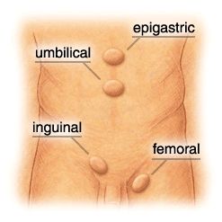
Hesselbach's triangle - Inguinal triangle


Ilioinguinal nerve - referred pain


Genitofemoral Nerve

  1. In human anatomy, the genitofemoral nerve originates from the upper part of the lumbar plexus of spinal nerves. Its roots are L1 and L2 (lumbar).
  2. The genitofemoral nerve is responsible for both the efferent and afferentlimbs of the cremasteric reflex. The ilioinguinal nerve (L1) is also involved in the afferent limb of the reflex.
  3. It emerges on the anterior surface of the psoas major muscle and divides into two branches:
    1. The femoral branch, or lumboinguinal nerve, supplies skin anterior to the upper part of the femoral triangle
    2. The genital branch:

Obturator nerve

It descends through the fibers of the Psoas major, and emerges from its medial border near the brim of the pelvis; it then passes behind the common iliac vessels, and on the lateral side of the hypogastric vessels and ureter, which separate it from the ureter, and runs along the lateral wall of the lesser pelvis, above and in front of the obturator vessels, to the upper part of the obturator foramen.

Here it enters the thigh, through the obturator canal, and divides into an anterior and a posterior branch, which are separated at first by some of the fibers of the Obturator externus, and lower down by the Adductor brevis.

Femoral Nerve
?Dont have a specimen with internal??
Superior Gluteal Nerve - Waddling gate


    Minggu, 20 Februari 2011

    Left Femur and Patella

    Left Femur - upper end
    The pectineal line of the femur must not be confused with the pectineal line of the pubis, nor with the spiral line of the femur which is usually more prominent than the pectineal line.


    1. Fovea of the Head Os - Ligament of the head of the femur
    2. Greater Trochanter Os - Outside Anteriorly - Mm Gluteus Minimus, Top Mm Piriformis. On the inside of the great Trochanter, Gluteus medius and also MM Obturator Internus and Mm Gemelli. The large Quadraus Femoralis Mm. Inferior anterior the Mm Vastus Lateralis, 
    3. Head
    4. Intertrochanteric Line (anterior) - with the Iliofemoral Ligament. 
    5. Lesser Trochanter Os - with Psoas Major Mm attachment and the Mm Iliacus. Inferior from medial to lateral the Mm Vastus Medius. 
    6. Neck of the Femur
    7. Pectineal line
    8. Quadrate turbercle on the intertrochanteric crest
    9. Shaft - with Mm Vastus Intermedius.
    10. Spiral Line
    11. Trochanteric Fossa - Mm Obturator Externus 


    Left Femur - Shaft

    1. Gluteal Tuberosity Os - Mm Gluteus Maximus
    2. Lateral Supracondylar line
    3. Lesser Throchanter
    4. Linea Aspera. The rough linea aspera oftern shows a distinct medial and lateral lips; the lateral lip continues upward as the gluteal tuberosity. Muscle attachments from medial to lateral: Mm Vastus Medialis, Adductor Longus Mm, Adductor Brevis, Mm Adductor Magnus, Short Head of Biceps Femoralis Mm, Vastus Lateralis Mm, Vastus Intermedius.  
    5. Medial Supracondylar Line
    6. Pectineal Line - Mm Pectineus Muscle. 

    Left Patella


    Left Femur - Lower end


    Ya-Khâliq (The Creator,  The Planner) 


     - Oh how great the creator of the heavens and earth must be if he creates femurs with such precision! the One who continues to plan, measure out and create, and who has the power to change things from non-existing to existing.
    JEHOVAH BORE = The Lord Creator (Is. 40:28)

    Sabtu, 19 Februari 2011

    Brachial Plexus


    Brachial Plexus (1/3)


    Brachial Plexus (2/3)


    Brachial Plexus (3/3)


    Upper arm muscles: Attachments


    Upper arm muscles: Attachments (part 1/6)


    Upper arm muscles: Flexors (part 2/6)


    Upper arm muscles: Extensors (part 3/6)


    Forearm muscles: Attachments (part 4/6)


    Forearm muscles: Flexors (part 5/6)


    Forearm muscles: Extensors (part 6/6)