Explanation
On the labeled CT image, C is the top of the main pulmonary artery. The structure labeled A is the superior vena cava, with the azygos vein entering from posteriorly. The structure labeled B is the ascending aorta. The structure labeled D is the descending or thoracic aorta. The vessels that connect directly to the heart are called collectively the 'great vessels', while the next set of branches and tributaries beyond this supplying the head and upper extremity are termed 'brachiocephalic vessels'.
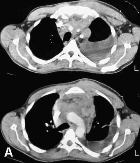
Image A shows two images from the CT of a patient with weight loss and fatigue. The same two images are shown with labels on key structures in Image B.
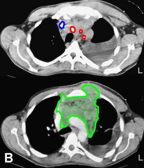
The three branches from the aortic arch (brachiocephalic, left common carotid, and left subclavian) are outlined in red, and the right brachiocephalic vein is outlined in blue. The left brachiocephalic vein is obliterated. A large mass (outlined in green) infiltrates the anterior mediastinum, surrounding vessels and growing into the left parasternal region. At biopsy, this was an aggressive lymphoma.
Tidak ada komentar:
Posting Komentar