Tampilkan postingan dengan label Pulmonary Trunk. Tampilkan semua postingan

Minggu, 05 Juni 2011

Pulmonary Artery or Pulmonary Trunk


eled CT:  

labeled CT: 

Explanation

The right and left clavicles are outlined in blue.  They are positioned obliquely for a chest CT scan because the patient's arms are raised overhead.  This rotates the scapulae and lifts the lateral ends of the clavicles from the usual resting position. The patient's arms are overhead for this study because it decreases artifacts in the upper portion of the chest due to excessive bone.  If patients are scanned with their arms at their sides, the course of the beam in the coronal plane must pass through the humeri, scapulae, and spine, and this produces streak artifact.

Explanation

The labeled CT shows the main pulmonary artery and its two branches, the right and left pulmonary arteries outlined in blue.


Figure A shows an abnormality of the right pulmonary artery.
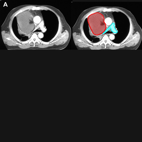
created on: 06/21/09
The main, right and left pulmonary arteries are outlined in blue. There is a large right mediastinal mass of relatively low attenuation (a liposarcoma) that is compressing the right pulmonary artery.

Figure B shows a different abnormality of the pulmonary artery.
created on: 06/21/09
In this case, a small cell lung carcinoma (outlined in red) has directly invaded the right pulmonary artery  (outlined in purple), producing complete obstruction of flow to the right lung.  Only bronchial artery branches remain patent to supply blood to the right lung tissues.

Selasa, 31 Mei 2011

Pulmonary Artery or Pulmonary Trunk


Explanation

On the labeled CT image, C is the top of the main pulmonary artery.  The structure labeled A is the superior vena cava, with the azygos vein entering from posteriorly.  The structure labeled B is the ascending aorta.  The structure labeled D is the descending or thoracic aorta.  The vessels that connect directly to the heart are called collectively the 'great vessels', while the next set of branches and tributaries beyond this supplying the head and upper extremity are termed 'brachiocephalic vessels'.
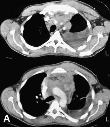
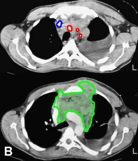
Image A shows two images from the CT of a patient with weight loss and fatigue.  The same two images are shown with labels on key structures in Image B.
created on: 07/27/09
The three branches from the aortic arch (brachiocephalic, left common carotid, and left subclavian) are outlined in red, and the right brachiocephalic vein is outlined in blue.  The left brachiocephalic vein is obliterated.  A large mass (outlined in green) infiltrates the anterior mediastinum, surrounding vessels and growing into the left parasternal region.  At biopsy, this was an aggressive lymphoma.