eled CT:
labeled CT:
Explanation
The right and left clavicles are outlined in blue. They are positioned obliquely for a chest CT scan because the patient's arms are raised overhead. This rotates the scapulae and lifts the lateral ends of the clavicles from the usual resting position. The patient's arms are overhead for this study because it decreases artifacts in the upper portion of the chest due to excessive bone. If patients are scanned with their arms at their sides, the course of the beam in the coronal plane must pass through the humeri, scapulae, and spine, and this produces streak artifact.
Explanation
The labeled CT shows the main pulmonary artery and its two branches, the right and left pulmonary arteries outlined in blue.
Figure A shows an abnormality of the right pulmonary artery.
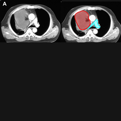
The main, right and left pulmonary arteries are outlined in blue. There is a large right mediastinal mass of relatively low attenuation (a liposarcoma) that is compressing the right pulmonary artery.
Figure B shows a different abnormality of the pulmonary artery.

In this case, a small cell lung carcinoma (outlined in red) has directly invaded the right pulmonary artery (outlined in purple), producing complete obstruction of flow to the right lung. Only bronchial artery branches remain patent to supply blood to the right lung tissues.
Tidak ada komentar:
Posting Komentar