labeled CT:
unlabeled CT:
Explanation
The labeled CT shows the main pulmonary artery and its two branches, the right and left pulmonary arteries outlined in blue.
Figure A shows an abnormality of the right pulmonary artery.
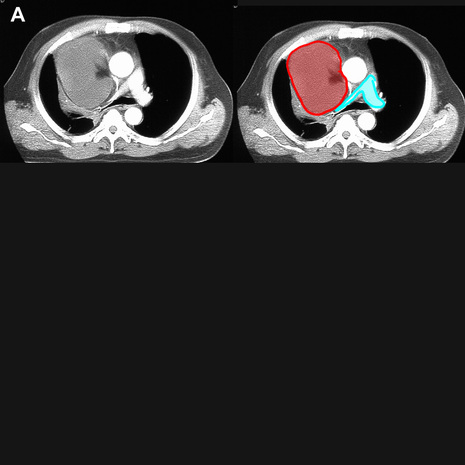
The main, right and left pulmonary arteries are outlined in blue. There is a large right mediastinal mass of relatively low attenuation (a liposarcoma) that is compressing the right pulmonary artery.
Figure B shows a different abnormality of the pulmonary artery.

In this case, a small cell lung carcinoma (outlined in red) has directly invaded the right pulmonary artery (outlined in purple), producing complete obstruction of flow to the right lung. Only bronchial artery branches remain patent to supply blood to the right lung tissues.
Tidak ada komentar:
Posting Komentar