labeled CT:
unlabeled CT:
Explanation
The labeled CT images show the course of the ascending aorta, aortic arch, and descending aorta, along with the three major branches that arise from the aortic arch. The branch outlined in blue is the brachiocephalic artery.
The aorta is outlined in red, the left common carotid artery in purple, and the left subclavian artery in yellow. The only branches that arise from the ascending aorta are the coronary arteries. The descending aorta gives off the bronchial arteries, the esophageal arteries, mediastinal arteries and the posterior intercostal arteries. After the thoracic aorta enters the abdomen via the aortic hiatus of the diaphragm, it becomes the abdominal aorta. Two of the midline branches of the upper abdominal aorta are also labeled: the celiac artery (in green) and the superior mesenteric (in dark blue).
Image A shows an abnormality of the branches off the aorta:
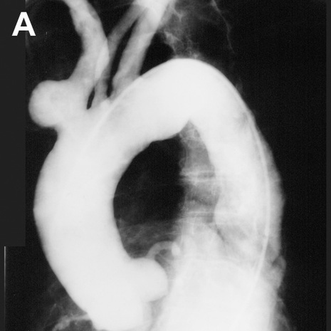
Image B shows the aortic arch and its branches outlined:
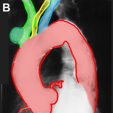
The aorta is outlined in red. The first branch, the brachiocephalic artery, is shown in green. There is a rounded outpouching near the origin of this vessel that protrudes from the anterior side of the vessel. A contour abnormality of this type is called a saccular aneurysm, and in this patient this abnormality was visible on the lateral chest radiograph, simulating an anterior mediastinal mass. The normal left common carotid artery is shown in yellow and the normal left subclavian artery is shown in blue.
Tidak ada komentar:
Posting Komentar