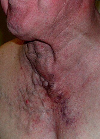
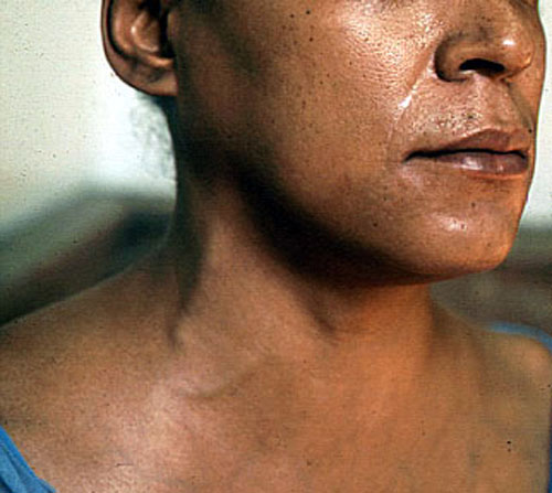
The third part of the subclavian artery. The subclavian artery has 3 parts - separated by the scalenus anterior in thirds - the last third is the part between the scalenus anterior and the clavicle. It is a fingerbreath superior to the clavicle.
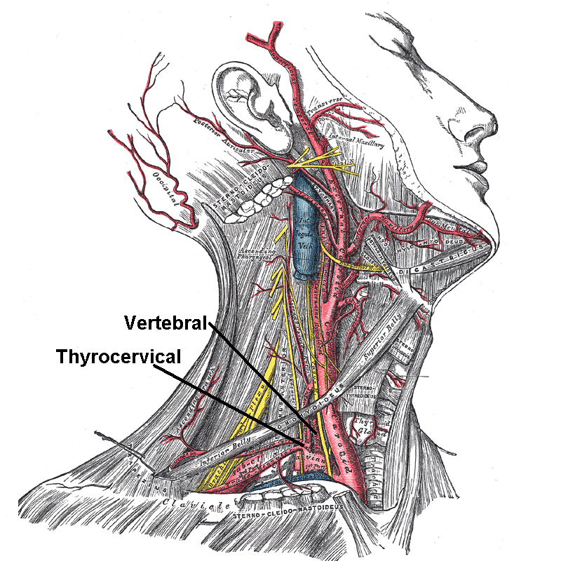

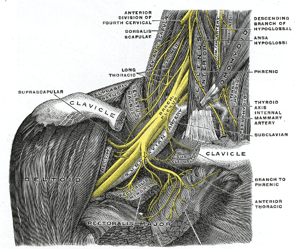
The transverse cervical artery arises from the thyrocervical trunk - a branch of the subclavian artery. The transverse cervical artery runs superficially and laterally across the posterior triangle, 2 to 3 cm superior to the clavicle.
The Suprascapular artery from the branch of the thyrocervical trunk - runs posterior to the clavical to the muscles around the scapula.
| Artery: Transverse cervical artery | |
|---|---|
 | |
| Superficial dissection of the right side of the neck, showing the carotid and subclavian arteries. Transverse cervical artery is labeled, branching from the thyrocervical_trunk. | |
 | |
| Superficial and deep branches from the transverse cervical artery. | |
| Latin | arteria transversa cervicis, arteria transversa colli |
| Gray's | subject #148 82 |
| Supplies | Trapezius Sternocleidomastoid |
| Source | Thyrocervical trunk |
| Branches | Superficial branch Deep branch |
| Vein | Transverse cervical veins |


Nerves in the posterior triangle. The Accessory nerve (CN XI) that divides the posterior triangle into a superior and inferior parts. The Accessory nerve is a motor nerve consisting of spinal (C1 to C5) and cranial roots. The spinal roots travel superiorly and enter the posterior cranial fossa through the foramen magnum. Here they join the cranial roots of the accessory nerve from the Medulla. Both combined roots leave the skull through the jugular foramen.
The Spinal roots separates immediately and supplies the Sternocleidomastoid muscle and the trapezius muscle.
Lesions to the Accessory Muscle are rare. The Accessory Nerve may be damaged by Traumatic injury, tumours to the base of the skull, fractures involving the jugular foramen, and neck lacerations. Presents with weakness in turning the head to the OPPOSITE site against resistance. Unilateral paralysis of the trapezius muscle is evident by the patients inability to elevate and retract the shoulder, and is unable to lift the arm above the horizontal level. Drooping of the shoulder is obvious.
The Accessory Nerve function can also be functionally impaired by inflamed lymph nodes in the neck. During surgical dissection of the neck lymph nodes care should be taken to isolate and preserve the Accessory Nerve.

| Nerve: Accessory nerve | |
|---|---|
 | |
| Plan of upper portions of glossopharyngeal, vagus, and accessory nerves. | |
 | |
| Inferior view of the human brain, with the cranial nerves labelled. | |
| Latin | nervus accessorius |
| Gray's | subject #206 913 |
| Innervates | sternocleidomastoid muscle, trapezius muscle |
| MeSH | Accessory+Nerve |
The Cervical Plexus
, Anterior triangle, Arteries, Carotid Artery, Carotid Body, , Hyoid Muscles, Muscles, Neck, Nerves, , Posterior triangle, raised JVP, , XI