Sternocleidomastoid Muscle.
Sternocleidomastoid | |
|---|---|
 | |
The triangles of the neck. (Anterior triangles to the left; posterior triangles to the right.) | |
 | |
Muscles of the neck. Lateral view. | |
musculus sternocleidomastoideus | |
mastoid process of the temporal bone, superior nuchal line of the occipital bone | |
motor: accessory nerve sensory: cervical plexus (ventral ramus of the second cranial nerve) | |
Acting alone, tilts head to its own side and rotates it so the face is turned towards the opposite side. Acting together, flexes the neck, raises the sternum and assists in forced inspiration. | |
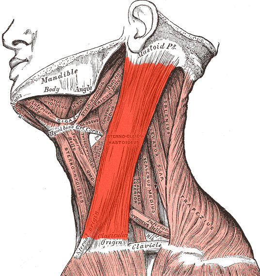 .
.The Sternocleidomastoid is crossed by the platysma and the External Jugular Vein. The Sternocleidomastoid covers the great vessels of the neck and the cervical plexus of the nerves. 


Congenital torticollis: The Sternocleidomastoid may be injured at birth resulting in congenital torticollis. There is fixed rotation and tilting of the head owing to contracture of the sternocleidomastoid muscle. Birth injury, tearing fibers of the muscle during NVD vaginal delivery - bleeding - swelling and then fibrosis of the sternocleidomastoid. This shortening of the muscle is evident at age 3 to 4 when the neck elongates. Managed by physiotherapy that promotes daily stretching and tilting the head.
Spasmodic torticollis may develop in adults - Stiff neck.

Posterior Triangle of the Neck.
The Sternocleidomastoid, the Trapezius and Clavicle form the posterior triangle of the neck. The trapezius and the Sternocleidomastoid meet at the superior nuchal line - where the Occipital Artery passes through the apex of the posterior triangle, before it ascends over the posterior aspect of the head.
The roof of the Posterior triangle of the Neck - Covered by deep fascia and superficial to this fascia is the Platysma veins, nerves and skin.


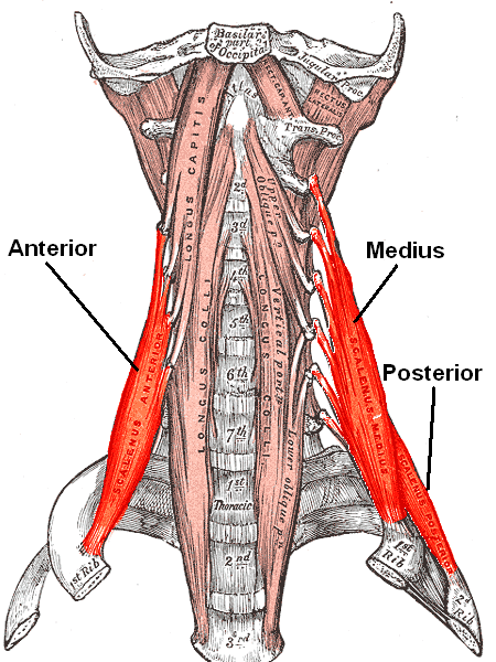
The muscular FLOOR of the posterior triangle is formed by splenius capitus, levator scapulae, scalenus medius and scaleneus posterior muscles. The four mascles are covered by a carpet of Deep Cervical Fascia. The Spenius capitus muscle arises from the the ligamentum nuchae and the spinous process of the superior thoracic vertebrae. The Spenius Capitus runs superolaterally and inserts into the mastoid process.
Levator scapulae muscle lifts the scapula, arises from the first 4 cervical vertebrae and inserts into the medial boarder of the scapulae.
Scalenus Posterior Muscle arises from the lower two to three cervical vertebrae (4th, 5th, 6th cervical vertebrae) and inserts into the 2nd rib.
Scalenus Medius Muscle arises from the posterior tubercles of ALL the cervical vertebrae. Inserts on the posterior part of the 1st rib. The Scalenus medius lies posterior to the roots of the brachial plexus.
Levator scapulae muscle lifts the scapula, arises from the first 4 cervical vertebrae and inserts into the medial boarder of the scapulae.
Scalenus Posterior Muscle arises from the lower two to three cervical vertebrae (4th, 5th, 6th cervical vertebrae) and inserts into the 2nd rib.
Scalenus Medius Muscle arises from the posterior tubercles of ALL the cervical vertebrae. Inserts on the posterior part of the 1st rib. The Scalenus medius lies posterior to the roots of the brachial plexus.


| Levator scapulae muscle | |
|---|---|
 | |
| Muscles connecting the upper extremity to thevertebral column. (Levator scapula visible at upper right, at the neck.) | |
| Latin | musculus levator scapulae |
| Gray's | subject #121 435 |
| Origin | Posterior tubercles oftransverse processes of C1 - C4 vertebrae |
| Insertion | Superior part of medial border of scapula |
| Artery | dorsal scapular artery |
| Nerve | cervical nerve (C3, C4) anddorsal scapular nerve (C5) |
| Actions | Elevates scapula and tilts itsglenoid cavity inferiorly by rotating scapula |
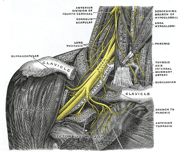
Contents of the Posterior Triangle: Vessels and nerves of the Neck and Upper Limb'
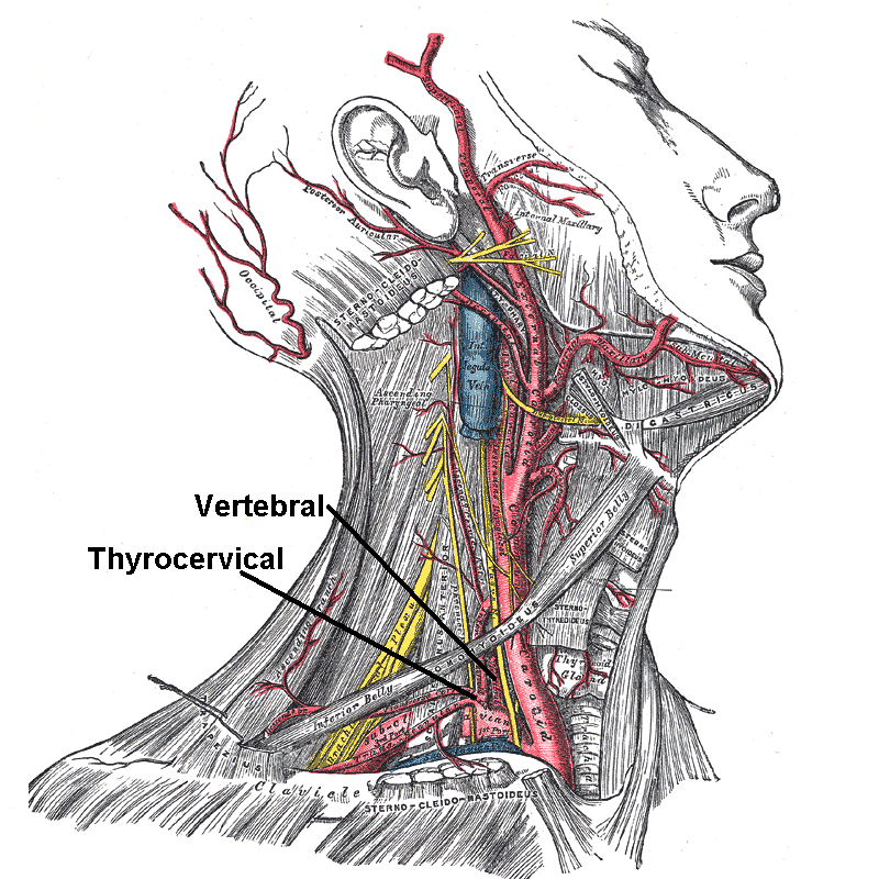
The External Jugular Vein begins near the angle of the mandible, inferior to the lobule of the auricle, by union of the posterior division of the retromandibular vein and the posterior auricular vein. The external jugular vein crosses the sternocleidomastoid muscle in the superficial fascia and then pierces the fascia roof of the posterior triangle at he posterior boarder of the sternocleidomastoid muscle. It then passes obliquely through the inferior part of the posterior triangle to empty in the subclavian vein 2cm superior to the clavicle.
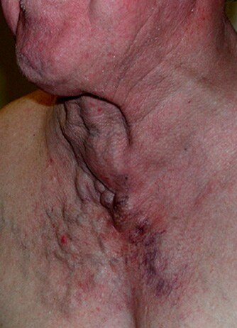
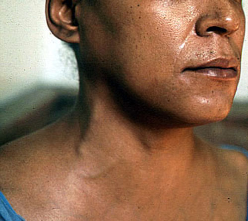
A raised JVP in heart failure may be seen throughout the course of the external jugular Vein. A JVP is raised in increased thoracic pressure, obstructed by a tumour, and enlarged supraclavicular lymph nodes. Opera singers have bilateral enlargement of their external jugular veins due to prolonged periods of increased intrathoracic pressure. IV Fluid overload may have a engorged JVP. Laceration of the external jugular about 5cm superior to the clavicle, may result in air embolism as the External Jugular can not retract when it pierces the deep fascia of the posterior triangle - the patient may present with dyspnea and cyanosis.
| Vein: External jugular vein | |
|---|---|
 | |
| Veins of the head and neck. (External jugular visible at center.) | |
 | |
| Veins | |
| Latin | vena jugularis externa |
| Gray's | subject #168 646 |
| Drains from | cranium, face |
| Source | posterior facial vein,posterior auricular vein,anterior jugular vein |
| Drains to | subclavian vein |
| MeSH | Jugular+Veins |