Selasa, 31 Mei 2011

emergency surgical airway be inserted

(11) You and your chief resident are the first responders to a code green (respiratory arrest) on the thoracic surgery ward. Amid the mayhem which follows, your chief resident tells you to perform an emergency surgical airway on the non-responsive patient while he looks for a code cart on another floor. Through which anatomic structure should the emergency surgical airway be inserted? 


Explanation

Correct Answer: 
Crico-thyroid membrane
Take-Home Message: 
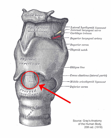
Emergency access to the airway can most safely be obtained by piercing the crico-thyroid membrane in the midline of the neck. This is termed a cricothyroidotomy.
Explanation of Correct Answer: 
The cricoid cartilage is the only tracheal cartilage that is a complete ring. It is located immediately inferior to the wide,flat thyroid cartilage. There is generally a palpable indentation between the thyroid and cricoid cartilages, which represents the crico-thyroid membrane. These surface landmarks make this location an ideal one for performing emergency surgery to gain access to the airway. In the midline, there are no large crossing vessels and no important nerves.
Explanation of Incorrect Answers: 
The 
hypopharynx is not an appropriate location for emergency airway access.
The 
thyroid cartilage is too hard to penetrate for placement of emergency airway access and might lead to laryngeal damage.
The 
cricoid cartilage is too hard to penetrate for placement of emergency airway access.
The 
tracheal rings limit the ability to place emergency airways.
Clinical Pearls: 
If a patient needs prolonged respiratory support on a ventilator, a formal tracheostomy (or tracheotomy) procedure may be performed. This is different from the cricothyroidotomy outlined above in that the tracheostomy tube is placed through the tracheal rings below the cricoid cartilage. A tracheostomy is not a good emergency procedure because the thyroid is in front of the trachea and must be divided to place a tracheostomy.
For more information on this topic, please click on the following link(s): 
(While these web sources have been vetted by our content experts, please use them with caution --- the peer-reviewed literature should be the ultimate source of medical information.) 
Cricothyroidotomy.
 http://en.wikipedia.org/wiki/Cricothyroidotomy 
Tracheotomy.
 http://en.wikipedia.org/wiki/Tracheostomy 
created on: 03/23/09

painless swelling of his left scrotum

(10) A 63-year old man recently-diagnosed with kidney cancer presents to your clinic reporting the new onset of painless swelling of his left scrotum. On examination, you find that the swelling is caused by engorged spermatic veins around the left testis. His right testis and scrotum are normal. This new physical finding suggests that his kidney cancer has spread to the: 


Explanation

Correct Answer: 
Renal vein
Take-Home Message: 
The venous drainage of the right and left testes is different (outlined in yellow in the image below): the left testis drains into the left renal vein while the right testis drains directly into the IVC. Obstruction of the left renal vein (e.g. from a renal tumor) can cause rapid engorgement of the spermatic veins around the left testis (termed a varicocele).
Explanation of Correct Answer: 
The testes receive their arterial supply from the gonadal arteries, which ariase from the anterior surface of the abdominal aorta near its midpoint. The right gonadal vein, which drains the testis and its coverings, travels up from the scrotum through the inguinal canal and into the abdominal cavity in company with the right gonadal artery, draining into the inferior vena cava near the level of the renal vessels. However, the left gonadal vein has a slightly different course, initially traveling with the gonadal artery but draining into the left renal vein rather than the cava. So, if a tumor involves the left renal vein, as renal cell carcinomas may sometimes do, this will typically block the drainage from the left testis and scrotum, causing engorgement of the veins in this area.
Explanation of Incorrect Answers: 
Spread of tumor to the 
abdominal aorta could compromise the gonadal artery but would not cause backup of venous blood in the scrotum.
Spread to the 
spermatic artery would not result in enlarged vessels in the scrotum, but might cause testicular infarction.
Spread to the 
inferior vena cava would be unlikely to block selectively the left gonadal venous drainage although it might block drainage of the right testis which drains into the IVC, depending on the location involved.
Spread to the 
inferior mesenteric vein would likely be asymptomatic, or might cause GI symptoms but would not interfere with venous drainage from the scrotum.
Spread to the 
testis would not cause venous blockage.
Clinical Pearls: 
(1) Lymphatic drainage of the testis follows venous drainage, so that metastases to nodes from testicular carcinomas first appear near the level of the renal vessels, where both gonadal veins drain (on the right directly into the IVC and on the left into the left renal vein).
(2) If a unilateral left varicocele develops in one of your patients, a left renal tumor needs to be ruled out by radiologic imaging.
For more information on this topic, please click on the following link(s): 
(While these web sources have been vetted by our content experts, please use them with caution --- the peer-reviewed literature should be the ultimate source of medical information.) 
http://www.emedicine.com/radio/topic601.htm 
Reference: 
Curti BD. Renal cell carcinoma. JAMA 2004; 292:97-100. 
created on: 03/23/09

Azygos vein

(09) A 41-year old woman develops superior vena cava syndrome due to a tumor located at the junction of the right and left brachiocephalic veins. What vein can still provide direct inflow into her superior vena cava? 

Explanation

Correct Answer: 
Azygos vein
Take-Home Message: 
The azygos vein (arrow 1, below) can provide an important collateral pathway for return of blood to the heart in cases of superior vena cava (SVC) obstruction [also known as SVC syndrome].
Explanation of Correct Answer: 
The azygos and hemiazygos system of veins are located in the posterior mediastinum and provides an important potential collateral pathway for blood when there is either blockage of the superior or inferior vena cava. Since these veins do not contain valves, blood can easily flow in either direction. If there is obstruction of the superior vena cava or brachiocephalic veins superior to the inflow of the azygos, blood from the upper body can enter the azygos system from anterior to posterior intercostal veins and still reach the superior vena cava below the point of obstruction via the azygos arch. If the obstruction is below the entry point of the azygos into the superior vena cava, this same system of veins can still return blood to the heart from below via connections of the azygos to the inferior vena cava or its abdominal tributaries.
Explanation of Incorrect Answers: 
The 
internal jugular vein joins the subclavian veins to return blood flow into the brachiocephalic veins. This vein does not provide a route for blood to bypass the blockage at the junction of the brachiocephalic veins.
The 
external jugular vein joins the subclavian vein more laterally, combining with the internal jugular veins to form the brachiocephalic veins. This vein does not provide a route for blood to bypass the blockage at the junction of the brachiocephalic veins.
The 
internal mammary vein returns blood flow via the brachiocephalic trunk. This vein does not provide a route for blood to bypass the blockage at the junction of the brachiocephalic veins.
Clinical Pearls: 
Non-small cell (bronchogenic) cancer is the major cause of malignant SVC syndrome, while histoplasmosis is the most common cause of benign SVC syndrome.
For more information on this topic, please click on the following link(s): 
(While these web sources have been vetted by our content experts, please use them with caution --- the peer-reviewed literature should be the ultimate source of medical information.) 
http://www.emedicine.com/emerg/topic561.htm 
http://en.wikipedia.org/wiki/Azygos_vein 

created on: 03/23/09

gag reflex


(06) A 12-year old boy has a very active gag reflex when throat cultures are attempted. The EFFERENT limb of this reflex is carried in what cranial nerve? (please select the best answer)   

Choices

  •  Trigeminal
  •  Vagus
  •  Glossopharyngeal
  •  Facial
  • Explanation

    Correct Answer: 
    Vagus (efferent)
    Take-Home Message: 
    The gag reflex results from sensory innervation through the glossopharygeal nerve (afferent limb) and motor innervation through the vagus nerve (efferent limb).
    Explanation of Correct Answer: 
    The soft palate and surrounding regions of the oropharynx are richly supplied with nerve endings, which carry sensory signals to the spinal cord by the glossopharyngeal nerve (afferent limb). Even light tough applied to these regions may elicit a strong gag reflex in some people. The efferent limb of this reflex is carried by the vagus nerve, which supplies the pharyngeal constrictors, levator veli palatini, palatoglossus, palatopharyngeus and the laryngeal muscles.
    Explanation of Incorrect Answers: 
    The 
    glossopharyngeal nerve carries afferent fibers for the gag reflex, but not efferent fibers. The only muscle that receives motor innervation from the glossopharyngeal is the stylopharyngeus muscle, which does not play a role in the gag reflex.
    The 
    trigeminal nerve is the major sensory nerve of the face and supplies motor fibers to the muscles of mastication, but not the muscles involved in the gag reflex.
    The 
    facial nerve is the major motor nerve of the muscles of facial expression, as well as stylohyoid, stapedius and posterior belly of digastric, none of which are involved in the gag reflex
    Clinical Pearl: 
    The gag reflex can be impaired in patients after stroke and can lead to increased risk of aspiration pneumonia.
    For more information on this topic, please click on the following link(s): 
    (While these web sources have been vetted by our content experts, please use them with caution --- the peer-reviewed literature should be the ultimate source of medical information.) 
    http://library.med.utah.edu/neurologicexam/html/cranialnerve_anatomy.html#12 
    http://www.neuroanatomy.wisc.edu/virtualbrain/BrainStem/08CNX.html 

1st Rib


created on: 06/09/09
Which of the labeled structures on this single CT image represents the first rib?

Explanation

The area indicated by A is the region of the left clavicular head. The area indicated by B is the left first rib.  The area indicated by C is the left second rib.  The area indicated by D is the left T2 transverse process.  
For comparison, a common abnormality of the upper ribs is show below.  
Image A is a radiograph of the cervical spine showing additional tiny ribs originating from the 7th cervical vertebra, called cervical ribs.  These ribs are often asymptomatic, but can press on vessels or nerves.  The extra ribs are shown in red.  The normal first ribs are shown in blue and the 7th cervical and first thoracic vertebra in yellow.  
created on: 06/09/09

anterior scalene muscle


labeled movie:
 

Explanation

The area outlined in red represents the anterior scalene muscle, which extends from the cervical spine to the upper ribs.  It is an important CT landmark for the structures at the thoracic apex.  The brachial plexus emerges posterior to it, and the subclavian vein (outlined in blue) passes anterior to it, and the subclavian artery (outlined in yellow) passes posterior to it.
Image A below shows an abnormality on coronal T1-weighted MR in the region of the anterior scalenes.  Image B shows the same CT with structures outlined.

created on: 07/23/09

created on: 07/23/09
This patient has a lung cancer that has metastasized to C7 and is growing out to the right, invading the region of the brachial plexus and causing arm pain and weakness.  The tumor is shown in red.  The middle and posterior scalene muscles, which pass posterior to the brachial plexus, are outlined in yellow.  The bracheal plexus is outlined in green.  The anterior scalenes are outlined in blue.

intercostal arteries


Explanation

The small structures outlined in red are the intercostal arteries, which come off the aorta (the larger structure outlined in red) at each level and travel just inferior to each rib.  Each intercostal artery travels together with an intercostal vein and an intercostal nerve.
There are most commonly 9 pairs of intercostal arteries that arise from the aorta, distributed to the T3-T11 levels.  Because the T12 branch is not between two ribs, it is called the subcostal artery.  The arteries that run between T1-T2 and T2-T3 most often arise from the costocervical trunk as the supreme (or highest, or superior) intercostal artery.  This is somewhat variable, and the arteries to these upper levels can arise from the aorta or from other vessels such as the vertebral artery.
Image A is an unlabeled image from a T1-weighted sagittal MR of a patient with abnormalities of the intercostal arteries.
created on: 07/17/09

Image B shows the abnormal aorta, outlined in red, which has a marked narrowing just below the level of the ductus:  a coarctation.  In this condition, the intercostal arteries may become markedly enlarged as they serve as a collateral pathway for blood to get past the area of narrowing, as shown here.
created on: 07/17/09

1st Rib


Explanation

The yellow outline in the movie indicates the location of the left first rib.  The left second rib is indicated in pink.  

A clinical example is shown below of abnormal ribs for comparison.  
Image A shows an unlabeled CT image.
created on: 07/17/09
Image B shows the chondrosarcoma originating in an anterior left upper rib, outlined in red.  The mass contains considerable calcification, with a somewhat swirling shape that is characteristic of lesions arising from cartilaginous cells, shown in blue. 
created on: 07/17/09

Pulmonary Artery or Pulmonary Trunk


Explanation

On the labeled CT image, C is the top of the main pulmonary artery.  The structure labeled A is the superior vena cava, with the azygos vein entering from posteriorly.  The structure labeled B is the ascending aorta.  The structure labeled D is the descending or thoracic aorta.  The vessels that connect directly to the heart are called collectively the 'great vessels', while the next set of branches and tributaries beyond this supplying the head and upper extremity are termed 'brachiocephalic vessels'.
Image A shows two images from the CT of a patient with weight loss and fatigue.  The same two images are shown with labels on key structures in Image B.
created on: 07/27/09
The three branches from the aortic arch (brachiocephalic, left common carotid, and left subclavian) are outlined in red, and the right brachiocephalic vein is outlined in blue.  The left brachiocephalic vein is obliterated.  A large mass (outlined in green) infiltrates the anterior mediastinum, surrounding vessels and growing into the left parasternal region.  At biopsy, this was an aggressive lymphoma.

azygos vein


   Explanation

The azygos vein is outlined in orange.  It joins the superior vena cava from posteriorly and provides an important collateral pathway for blood return to the heart in cases of superior or inferior vena cava obstruction.  The superior vena cava is outlined in green.  It forms at the junction of the right and left brachiocephalic veins, which are indicated in blue.  
Image A and the accompanying movie show a different patient with an abnormality of the azygos.
created on: 07/13/09
The aorta is shown in red, the superior vena cava in dark blue, the azygos and other posterior collaterals in light blue and the top of the main pulmonary artery in purple.