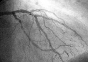
A coronary angiogram (an X-ray with radio-opaque contrast in the coronary arteries) that shows the left coronary circulation. The distal left main coronary artery(LMCA) is in the left upper quadrant of the image. Its main branches (also visible) are the left circumflex artery (LCX), which courses top-to-bottom initially and then toward the centre/bottom, and the left anterior descending (LAD) artery, which courses from left-to-right on the image and then courses down the middle of the image to project underneath of the distal LCX. The LAD, as is usual, has two large diagonal branches, which arise at the centre-top of the image and course toward the centre/right of the image.
One of most common angiograms performed is to visualize the blood in the coronary arteries. A long, thin, flexible tube called a catheter is used to administer the x-ray contrast agent at the desired area to be visualized. The catheter is threaded into an artery in the forearm, and the tip is advanced through the arterial system into the major coronary artery. X-ray images of the transient radiocontrast distribution within the blood flowing within the coronary arteries allows visualization of the size of the artery openings. Presence or absence of atherosclerosis or atheroma within the walls of the arteries cannot be clearly determined. See coronary catheterization for more detail..
[edit]Microangiography
[Microangiography] is commonly used to visualize tiny blood vessels.
[edit]Neuro-vascular angiography
Another increasingly common angiographic procedure is neuro-vascular digital subtraction angiography in order to visualise the arterial and venous supply to the brain. Intervention work such as coil-embolisation of aneurysms and AVM gluing can also be performed.
[edit]Peripheral Angiography
Main article: Peripheral artery occlusive disease
Angiography is also commonly performed to identify vessel narrowing in patients with leg claudication or cramps, caused by reduced blood flow down the legs and to the feet; in patients with renal stenosis (which commonly causes high blood pressure) and can be used in the head to find and repair stroke. These are all done routinely through the femoral artery, but can also be performed through the brachial or axillary (arm) artery. Any stenoses found may be treated by the use of atherectomy.
[edit]Other
Other angiographic uses include the diagnosis of retinal vascular disorders, such as diabetic retinopathy and macular degeneration.
[edit]Complications
[edit]Coronary Angiography
Coronary angiographys are common and major complications are rare. These include Cardiac arrhythmias, kidney damage, blood clots (which can cause heart attack or stroke),hypotension and pericardial effusion. Minor complications can include bleeding or bruising at the site where the contrast is injected, blood vessel damage on the route to the heart from the catheter (rare) and allergic reaction to the contrast.[1]
[edit]Cerebral Angiography
Major complications in Cerebral Angiography are also rare but include stroke, an allergic reaction to the anaesthetic other medication or the contrast medium, blockage or damage to one of the access veins in the leg, or thrombosis and embolism formation. Bleeding or bruising at the site where the contrast is injected are minor complications, delayed bleeding can also occur but is rare.[1]
Tidak ada komentar:
Posting Komentar