The great plexuses of the sympathetic are aggregations of nerves and ganglia, situated in the thoracic, abdominal, and pelvic cavities, and named the cardiac, celiac, and hypogastric plexuses. They consist not only of sympathetic fibers derived from the ganglia, but of fibers from the medulla spinalis, which are conveyed through the white rami communicantes. From the plexuses branches are given to the thoracic, abdominal, and pelvic viscera.

The Cardiac Plexus (Plexus Cardiacus)(Fig. 838).—The cardiac plexus is situated at the base of the heart, and is divided into a superficial part, which lies in the concavity of the aortic arch, and a deep part, between the aortic arch and the trachea. The two parts are, however, closely connected.
| The superficial part of the cardiac plexus lies beneath the arch of the aorta, in front of the right pulmonary artery. It is formed by the superior cardiac branch of the left sympathetic and the lower superior cervical cardiac branch of the left vagus. A small ganglion, the cardiac ganglion of Wrisberg, is occasionally found connected with these nerves at their point of junction. This ganglion, when present, is situated immediately beneath the arch of the aorta, on the right side of the ligamentum arteriosum. The superficial part of the cardiac plexus gives branches (a) to the deep part of the plexus; (b) to the anterior coronary plexus; and (c) to the left anterior pulmonary plexus. | 3 |
The deep part of the cardiac plexus is situated in front of the bifurcation of the trachea, above the point of division of the pulmonary artery, and behind the aortic arch. It is formed by the cardiac nerves derived from the cervical ganglia of the sympathetic, and the cardiac branches of the vagus and recurrent nerves. The only cardiac nerves which do not enter into the formation of the deep part of the cardiac plexus are the superior cardiac nerve of the left sympathetic, and the lower of the two superior cervical cardiac branches from the left vagus, which pass to the superficial part of the plexus.
The branches from the right half of the deep part of the cardiac plexus pass, some in front of, and others behind, the right pulmonary artery; the former, the more numerous, transmit a few filaments to the anterior pulmonary plexus, and are then continued onward to form part of the anterior coronary plexus; those behind the pulmonary artery distribute a few filaments to the right atrium, and are then continued onward to form part of the posterior coronary plexus.
| 5 | The left half of the deep part of the plexus is connected with the superficial part of the cardiac plexus, and gives filaments to the left atrium, and to the anterior pulmonary plexus, and is then continued to form the greater part of the posterior coronary plexus.
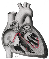
| 6 | The Posterior Coronary Plexus (plexus coronarius posterior; left coronary plexus) is larger than the anterior, and accompanies the left coronary artery; it is chiefly formed by filaments prolonged from the left half of the deep part of the cardiac plexus, and by a few from the right half. It gives branches to the left atrium and ventricle.
| 7 | | The Anterior Coronary Plexus (plexus coronarius anterior; right coronary plexus) is formed partly from the superficial and partly from the deep parts of the cardiac plexus. It accompanies the right coronary artery, and gives branches to the right atrium and ventricle. |
|
|
Tidak ada komentar:
Posting Komentar